Have you been diagnosed with breast cancer? Don’t fret! You need more imaging tests to confirm the accuracy of the disease! Your doctors will discuss with you which tests you need to identify any underlying symptoms of breast cancer. Imaging techniques utilize magnetic fields, x-rays, radioactive substances, and sound waves to generate pictures of the body’s interior.
Intermountain Medical Imaging conducts medical imaging tests to determine how far cancer might have spread, to look at the dubious areas that might be cancer, to search for probable cancer signs coming back after treatment, and to help decide on the fact which treatment is working. Below are the top 5 medical imaging techniques used to identify breast cancer in a patient.
- MRI
For women with a breast cancer diagnosis and those at high stake of breast cancer, MRI is the best strategy that can provide the most accurate and straightforward images of the breast. This radiation-free imaging technology creates 3D images of the breasts that doctors use in staging, screening, treatment response assessment, and pre-surgery planning.
MRI is performed on advanced 3 Testa scanners, which usually last approximately 45 minutes and need you to lie still for the most seamless results. These measures include warm blankets, eye covers, anti-anxiety treatments, and noise-canceling headphones.
- Mammography
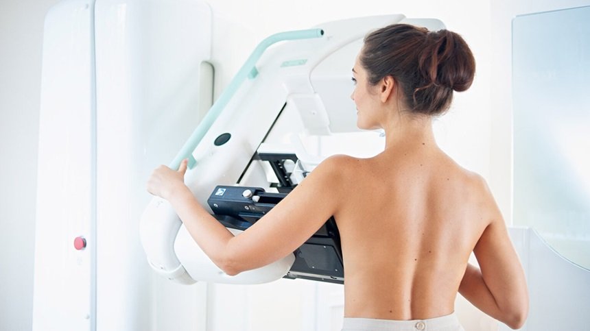
It uses X-rays to generate images of the breast, where the doctors perform screening mammograms for women without diagnostic mammograms and symptoms with concerned areas. The dedicated mammography technologists pay heed to the proper technique, breast compression, and body positioning to reduce your exposure to radiation during the mammogram.
A typical mammogram includes 3D mammography or tomosynthesis, which allows the radiologist to see through the thick tissue precisely with 2D mammography. This optimizes the scope of detecting cancer early and minimizes the risk of a false alarm.
- Ultrasound
Ultrasonography uses sound waves to generate an image on a video screen. A transducer, a tiny microphone-like instrument that emits sound waves, is moved over the skin surface and picks up the echoes as they rebound tissues. A PC turns these echoes into an image on the screen. It is possible to perform an ultrasound on the liver, underarms, or directly over the breast.
Several studies, including a large ACRIN trial assessing the utility of ultrasound screening in high-risk patients, have demonstrated that ultrasound can spot additional cancers in 3 to 4 of 1000 patients compared to MRI. Automated breast ultrasound machines have been manufactured to eliminate dependence on operators. Images can be reproducible and have 3D capability.
- Positron Emission Tomography or PET scan
For a PET scan, a radioactive type of sugar, FDG, penetrates the blood and gathers specifically in cancer cells. A PET scan is often merged with a CT scan using a unique tool that can do both simultaneously. This allows the doctor to compare the areas of higher radioactivity on the PET scan with an accurate snapshot on the CT scan.
- Image-Guided Breast Biopsy
It’s a minimally invasive process in which the doctors remove a sample of breast tissue from your breast. Proficient pathologists test the tissue under a microscope to look for cancerous cells. Moreover, they use the newest imaging technology to accurately pinpoint the biopsy area and guide the needle to a similar location. The shape, size, and tumor’s location determine the doctor’s biopsy strategy.
Conclusion
In this era of required radiation containment and cost, doctors must not conduct every test on every woman. It’s crucial that, along with establishing specificity, sensitivity, and other factors, these top 5 imaging techniques are highly beneficial for the early detection of breast cancer.


